
MIT CSAIL Cuts Placenta MRI Mapping Errors in Half With Neural Atlas
A new MIT method extends implicit shape learning into the organ interior, producing fold-free placenta atlases and clean voxel-wise MRI intensity maps in a single pass.
Takeaways
- MIT and Harvard team propose a volumetrically consistent implicit atlas for placenta MRI.
- Combines SDF reconstruction with a four-block Neural ODE diffeomorphic warp into a shared template.
- Adds Jacobian-determinant and biharmonic penalties evaluated on interior tetrahedral samples.
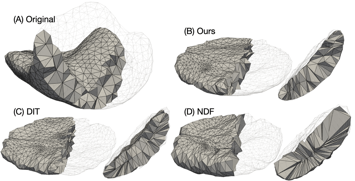
- Cuts tetrahedron flip rate to 4.04 percent versus 8.78 to 15.34 for DIT, DIF-Net, NDF.
- Enables single-pass voxel-wise intensity flattening preserving cotyledons and vascular structure.
- Trained on 111 in-vivo MRI scans from 78 pregnancies; no code released.
Building population atlases of the placenta is one of the ugliest problems in medical imaging. The organ drapes itself along the curved uterine wall in a different orientation for every patient, deforms with fetal motion and maternal breathing, and unlike the brain or heart, it has no widely accepted standardized geometric template. A team from MIT CSAIL, Massachusetts General Hospital, and Boston Children's Hospital has proposed a way to fix that using implicit neural representations and diffeomorphic flows. The trick is a set of penalties that finally force the network to behave sensibly inside the tissue, not just on its surface.
The paper, Volumetrically Consistent Implicit Atlas Learning, introduces a model that couples reconstruction of signed distance functions (SDFs) with neural diffeomorphic flow to learn a shared canonical template of the placenta. Jacobian-determinant and biharmonic penalties suppress local folding and promote globally coherent deformations. The model jointly reconstructs individual placentas, aligns them to a population-derived implicit template, and enables voxel-wise intensity mapping in a unified canonical space.
Why the interior of a shape is a blind spot
Deep implicit functions like DeepSDF represent a 3D object as a neural network that maps a coordinate to its signed distance from the surface. Template-based extensions such as DIT and DIF-Net add a learned warp field so that all shapes in a population get pushed into a shared canonical space, giving you correspondences across subjects. The problem, as the authors point out, is subtle but consequential: although losses are computed across the domain, the SDF loss gradient magnitude decays rapidly with distance from the zero-level set, leaving the deformation field under-constrained inside the volume.
In plain terms, the training signal only really pulls hard near the surface. The interior of the deformation field is free to fold, drift, or collapse without penalty. For a shape-classification task this is fine. For medical analysis of the placenta, where investigators want to compare