
Shanghai Jiao Tong's EMGauss Reconstructs Brain Slices 3dB Sharper Without Training Data
A Shanghai Jiao Tong team turns axial slice progression into a time axis and uses deformable Gaussian splatting to fill missing electron microscopy slices.
Takeaways
- EMGauss reconstructs isotropic 3D volumes from anisotropic electron microscopy stacks using dynamic Gaussian splatting.
- Axial slice index is treated as a time coordinate driving a deformation MLP over canonical 2D Gaussians.
- An EMA teacher-student loop generates pseudo-labels on unseen slices to prevent overfitting on sparse supervision.
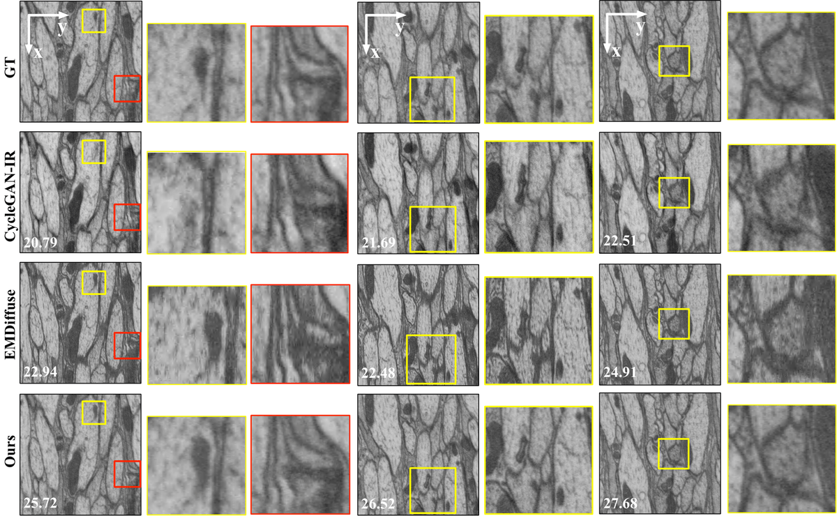
- Beats EMDiffuse, CycleGAN-IR, and IsoVEM by roughly 2.5-3 dB PSNR on EPFL and FIB-25 benchmarks.
- Enables continuous slice synthesis at arbitrary depths and improves SAM2 downstream segmentation IoU to 0.969.
- Full paper on arXiv; no official code release yet.
Volume electron microscopy can resolve neural tissue at nanometer scale, but scanning a specimen slice by slice creates a persistent headache: the lateral (xy) resolution is much sharper than the depth (z) resolution, producing squashed, anisotropic volumes. A group from Shanghai Jiao Tong University has now proposed EMGauss, a self-supervised method that borrows the machinery of dynamic 3D Gaussian splatting to synthesize the missing slices in between, without relying on paired training data or the shaky assumption that biological structures look the same from any angle.
The impossible triangle
vEM workflows always trade one thing for another. Volume electron microscopy enables nanoscale 3D imaging of biological structures but remains constrained by acquisition trade-offs, leading to anisotropic volumes with limited axial resolution. Techniques like serial section SEM or SBF-SEM prioritize speed over z-fidelity, while methods that produce true isotropic voxels (such as enhanced FIB-SEM) only work on small volumes and require specialized instrumentation. So most large datasets ship with z-resolution six to ten times worse than xy.
Deep learning has been pressed into service to fix this. The common trick is to leverage the abundant high-resolution xy-plane images as a self-supervised reference, either by treating consecutive xy slices as video frames and interpolating between them, or by training a super-resolution network on xz/yz planes. Both tactics ride on a strong assumption: that local tissue looks statistically similar in every direction. Existing deep learning methods seek to restore isotropy by leveraging lateral priors, yet their assumptions break down for morphologically anisotropic structures. Neurons don't cooperate here. Elongated fibers and dendritic spines are directional by nature, so priors learned on xy planes leak artifacts when applied along z.